FRAGMENT:
According to the concept originally introduced by George D. Lundberg in the 1980s, the total testing process entails three essential and sequential parts, that are the preanalytical phase, the analytical phase and the postanalytical phase (1). Briefly, the preanalytical phase encompasses all those (prevalently) manually-intensive activities designed for obtaining, handling, transporting, preparing and storing biological samples before testing (2). Reliable evidence, accumulated after decades of research aimed to improve the total quality of the testing process, underpins the notion that the vast majority of problems in laboratory diagnostics are attributable to incorrect or inappropriate preanalytical activities (3).
REFERENCE:
Lippi, Giuseppe, and Camilla Mattiuzzi. “Biological Samples Transportation by Drones: Ready for Prime Time?” Annals of Translational Medicine 4.5 (2016): 92. PMC. Web. 6 Nov. 2017.
-----------------------------------------------------------
Sigue este Blog en Facebook y Twitter
Ayúdanos a traducir las fichas de seguridad de microorganismos.
| Lista de correo. Espere su aprobación. |
| Consultar este grupo |
lunes, 6 de noviembre de 2017
miércoles, 1 de noviembre de 2017
Molecular Viability Testing of UV-Inactivated Bacteria
The polymerase chain reaction (PCR) is effective at detecting bacterial DNA in samples, but it is unable to differentiate viable bacteria from inactivated cells or free DNA fragments. New PCR-based analytical strategies have been developed to address this limitation. Molecular viability testing (MVT) correlates bacterial viability with the ability to rapidly synthesize species-specific ribosomal RNA precursor (pre-rRNA) in response to brief nutritional stimulation. Previous studies demonstrated that MVT can assess bacterial inactivation by chlorine, serum, and low-temperature pasteurization. Here, we demonstrate that MVT can detect inactivation of Escherichia coli, Aeromonas hydrophila, and Enterococcus faecalis cells by ultraviolet (UV) irradiation. Some UV-inactivated E. coli cells transiently retained the ability to synthesize pre-rRNA post-irradiation (generating false-positive MVT results), but this activity ceased within one hour following UV exposure. Viable but transiently undetectable (by culture) E. coli cells were consistently detected by MVT. An alternative viability testing method, viability PCR (vPCR), correlates viability with cell envelope integrity. This method did not distinguish viable from UV-inactivated bacteria under some conditions, indicating that the inactivated cells retained intact cell envelopes. MVT holds promise as a means to rapidly assess microbial inactivation by UV treatment.
IMPORTANCE Ultraviolet (UV) irradiation is increasingly used to disinfect water, food, and other materials for human use. Confirming the effectiveness of UV disinfection remains a challenging task. In particular, microbiological methods that rely on rapid detection of microbial DNA can yield misleading results. This is due to the detection of “remnant” DNA associated with dead microbial cells. This report describes a novel method that rapidly distinguishes living from dead microbial cells after UV disinfection.
REFERENCE:
Kris M. Weigel, et al. Molecular Viability Testing of UV-Inactivated Bacteria. Appl Environ Microbiol. 2017 May 15; 83(10): e00331-17. Prepublished online 2017 Mar 10. Published online 2017 May 1. doi: 10.1128/AEM.00331-17. PMCID: PMC5411506
-----------------------------------------------------------
Sigue este Blog en Facebook y Twitter
viernes, 27 de octubre de 2017
#History 1990: Model for inactivation and disposal of infectious HIV and radioactive waste in a BL3 facility
A method is described for autoclaving low levels of solid infectious, radioactive waste. The method permits steam penetration to inactivate biologic waste, while any volatile radioactive compounds generated during the autoclave process are absorbed. Inactivation of radiolabeled infectious waste has been problematic because the usual sterilization techniques result in unacceptable radiation handling practices. If autoclaved under the usual conditions, there exists a high probability of volatilization or release of radioisotopes from the waste. This results in the radioactive contamination of the autoclave and the laboratory area where steam is released from the autoclave. Our results provide a practical method to inactivate and dispose of infectious radioactive waste. For our research, Bacillus pumilus spore strips and vaccinia virus were used as more heat-resistant surrogates of the human immunodeficiency virus (HIV). These surrogates were used because HIV is difficult to grow under most conditions and is less heat tolerant than the surrogates. In addition, B. pumilus has defined cell death values, whereas such values have not been established for HIV. Both B. pumilus and vaccinia virus are less hazardous to work with. The autoclave method is time efficient and can be performed by laboratory personnel with minimal handling of the waste. Furthermore, waste site handlers are able to visually inspect the solid waste containers and ascertain that inactivation procedures have been implemented.
-----------------------------------------------------------
Sigue este Blog en Facebook y Twitter
Ayúdanos a traducir las fichas de seguridad de microorganismos.
REFERENCE:
Stinson MC, et al. Model for inactivation and disposal of infectious human immunodeficiency virus and radioactive waste in a BL3 facility. Appl Environ Microbiol. 1990 Jan;56(1):264-8.-----------------------------------------------------------
Sigue este Blog en Facebook y Twitter
Ayúdanos a traducir las fichas de seguridad de microorganismos.
miércoles, 25 de octubre de 2017
Dead or Alive: Molecular Assessment of Microbial Viability
Nucleic acid-based analytical methods, ranging from species-targeted PCRs to metagenomics, have greatly expanded our understanding of microbiological diversity in natural samples. However, these methods provide only limited information on the activities and physiological states of microorganisms in samples. Even the most fundamental physiological state, viability, cannot be assessed cross-sectionally by standard DNA-targeted methods such as PCR. New PCR-based strategies, collectively called molecular viability analyses, have been developed that differentiate nucleic acids associated with viable cells from those associated with inactivated cells. In order to maximize the utility of these methods and to correctly interpret results, it is necessary to consider the physiological diversity of life and death in the microbial world. This article reviews molecular viability analysis in that context and discusses future opportunities for these strategies in genetic, metagenomic, and single-cell microbiology.
REFERENCE:
Cangelosi, Gerard A., and John S. Meschke. “Dead or Alive: Molecular Assessment of Microbial Viability.” Ed. H. L. Drake. Applied and Environmental Microbiology 80.19 (2014): 5884–5891. PMC. Web. 4 Sept. 2017.
-----------------------------------------------------------
Sigue este Blog en Facebook y Twitter
REFERENCE:
Cangelosi, Gerard A., and John S. Meschke. “Dead or Alive: Molecular Assessment of Microbial Viability.” Ed. H. L. Drake. Applied and Environmental Microbiology 80.19 (2014): 5884–5891. PMC. Web. 4 Sept. 2017.
Nucleic acid-based analytical methods, ranging from species-targeted PCRs to metagenomics, have greatly expanded our understanding of microbiological diversity in natural samples. However, these methods provide only limited information on the activities and physiological states of microorganisms in samples. Even the most fundamental physiological state, viability, cannot be assessed cross-sectionally by standard DNA-targeted methods such as PCR. New PCR-based strategies, collectively called molecular viability analyses, have been developed that differentiate nucleic acids associated with viable cells from those associated with inactivated cells. In order to maximize the utility of these methods and to correctly interpret results, it is necessary to consider the physiological diversity of life and death in the microbial world. This article reviews molecular viability analysis in that context and discusses future opportunities for these strategies in genetic, metagenomic, and single-cell microbiology.
Sigue este Blog en Facebook y Twitter
lunes, 23 de octubre de 2017
Schrödinger’s microbes: Tools for distinguishing the living from the dead in microbial ecosystems
While often obvious for macroscopic organisms, determining whether a microbe is dead or alive is fraught with complications. Fields such as microbial ecology, environmental health, and medical microbiology each determine how best to assess which members of the microbial community are alive, according to their respective scientific and/or regulatory needs. Many of these fields have gone from studying communities on a bulk level to the fine-scale resolution of microbial populations within consortia. For example, advances in nucleic acid sequencing technologies and downstream bioinformatic analyses have allowed for high-resolution insight into microbial community composition and metabolic potential, yet we know very little about whether such community DNA sequences represent viable microorganisms. In this review, we describe a number of techniques, from microscopy- to molecular-based, that have been used to test for viability (live/dead determination) and/or activity in various contexts, including newer techniques that are compatible with or complementary to downstream nucleic acid sequencing. We describe the compatibility of these viability assessments with high-throughput quantification techniques, including flow cytometry and quantitative PCR (qPCR). Although bacterial viability-linked community characterizations are now feasible in many environments and thus are the focus of this critical review, further methods development is needed for complex environmental samples and to more fully capture the diversity of microbes (e.g., eukaryotic microbes and viruses) and metabolic states (e.g., spores) of microbes in natural environments.
REFERENCE:
Emerson JB1,et al. Schrödinger's microbes: Tools for distinguishing the living from the dead in microbial ecosystems. Microbiome. 2017 Aug 16;5(1):86. doi: 10.1186/s40168-017-0285-3.
-----------------------------------------------------------
Sigue este Blog en Facebook y Twitter
REFERENCE:
Emerson JB1,et al. Schrödinger's microbes: Tools for distinguishing the living from the dead in microbial ecosystems. Microbiome. 2017 Aug 16;5(1):86. doi: 10.1186/s40168-017-0285-3.
-----------------------------------------------------------
Sigue este Blog en Facebook y Twitter
jueves, 19 de octubre de 2017
Workplace Hazards to Reproduction and Development
This booklet contains information for those of you who are interested in identifying, evaluating, and reducing workplace reproductive and developmental health risks. The information provided ranges from descriptions of basic physiology and toxicology to specific guidance intended for health care providers, workplace health and safety personnel, workers, and employers.
REFERENCE:
Sharon L. Drozdowsky, B.S. and Stephen G. Whittaker, Ph.D. Hazards to Reproduction and Development: A Resource for Workers, Employers, Health Care Providers, and Health & Safety Personnel. Safety and Health Assessment and Research for Prevention (SHARP). Washington State Department of Labor and Industries. Technical Report Number: 21-3-1999
-----------------------------------------------------------
Sigue este Blog en Facebook y Twitter
martes, 17 de octubre de 2017
Laboratory-acquired infections of Salmonella enterica serotype Typhi in South Africa
BACKGROUND: Workers in clinical microbiology laboratories are exposed to a variety of pathogenic microorganisms. Salmonella species is among the most commonly reported bacterial causes of laboratory-acquired infections. We report on three cases of laboratory-acquired Salmonella enterica serotype Typhi (Salmonella Typhi) infection which occurred over the period 2012 to 2016 in South Africa.
METHODS: Laboratory investigation included phenotypic and genotypic characterization of isolates. Phenotypic analysis included standard microbiological identification techniques, serotyping and antimicrobial susceptibility testing. Genotypic analysis included the molecular subtyping methodologies of pulsed-field gel electrophoresis analysis, multilocus sequence typing and whole-genome sequencing (WGS); with WGS data analysis including phylogenetic analysis based upon comparison of single nucleotide polymorphism profiles of isolates.
RESULTS: All cases of laboratory-acquired infection were most likely the result of lapses in good laboratory practice and laboratory safety. The following critical issues were highlighted. There was misdiagnosis and misreporting of Salmonella Typhi as nontyphoidal Salmonella by a diagnostic laboratory, with associated public health implications. We highlight issues concerning the importance of accurate fluoroquinolone susceptibility testing and interpretation of results according to updated guidelines. We describe potential shortcomings of a single disk susceptibility screening test for fluoroquinolone susceptibility and suggest that confirmatory minimum inhibitory concentration testing should always be performed in cases of invasive Salmonella infections. These antimicrobial susceptibility testing issues resulted in inappropriate ciprofloxacin therapy which may have been responsible for failure in clearance of pathogen from patients. Salmonella Typhi capsular polysaccharide vaccine was not protective in one case, possibly secondarily to a faulty vaccine.
CONCLUSIONS: Molecular subtyping of isolates proved effective to investigate the genetic relatedness of isolates. Molecular subtyping data interpreted together with epidemiological data allowed us to pinpoint the most likely sources for our cases of laboratory-acquired infection.
REFERENCE:
Smith AM, et al. Laboratory-acquired infections of Salmonella enterica serotype Typhi in South Africa: phenotypic and genotypic analysis of isolates. BMC Infect Dis. 2017 Sep 29;17(1):656.
-----------------------------------------------------------
Sigue este Blog en Facebook y Twitter
lunes, 16 de octubre de 2017
Prevalence of murine leukemia virus contamination in human cell lines
Contaminations of cell cultures with microbiological organisms are well documented and can be managed in cell culture laboratories applying reliable detection, elimination and prevention strategies. However, the presence of viral contaminations in cell cultures is still a matter of debate and cannot be determined with general detection methods. In the present study we screened 577 human cell lines for the presence of murine leukemia viruses (MLV). Nineteen cell lines were found to be contaminated with MLV, including 22RV1 which is contaminated with the xenotropic murine leukemia virus-related virus variant of MLV. Of these, 17 cell lines were shown to produce active retroviruses determined by product enhanced reverse transcriptase PCR assay for reverse transcriptase activity. The contaminated cell lines derive from various solid tumor types as well as from leukemia and lymphoma types. A contamination of primary human cells from healthy volunteers could not be substantiated. Sequence analyses of 17 MLV PCR products and five complete MLV genomes of different infected cell lines revealed at least three groups of related MLV genotypes. The viruses harvested from the supernatants of infected cell cultures were infectious to uninfected cell cultures. In the course of the study we found that contamination of human genomic DNA preparations with murine DNA can lead to false-positive results. Presumably, xenotransplantations of the human tumor cells into immune-deficient mice to determine the tumorigenicity of the cells are mainly responsible for the MLV contaminations. Furthermore, the use of murine feeder layer cells during the establishment of human cell lines and a cross-contamination with MLV from infected cultures might be sources of infection. A screening of cell cultures for MLV contamination is recommended given a contamination rate of 3.3%.
-----------------------------------------------------------
Sigue este Blog en Facebook y Twitter
REFERENCE
Uphoff CC, Lange S, Denkmann SA, Garritsen HS, Drexler HG. Prevalence and characterization of murine leukemia virus contamination in human cell lines. PLoS One. 2015 Apr 30;10(4):e0125622. doi: 10.1371/journal.pone.0125622. eCollection 2015. PubMed PMID: 25927683; PubMed Central PMCID: PMC4416031.-----------------------------------------------------------
Sigue este Blog en Facebook y Twitter
viernes, 13 de octubre de 2017
Día mundial del lavado de manos, Octubre 15, 2017
miércoles, 11 de octubre de 2017
Triage and Management of Accidental Laboratory Exposures to Biosafety Level-3 and -4 Agents
The recent expansion of biocontainment laboratory capacity in the United States has drawn attention to the possibility of occupational exposures to BSL-3 and -4 agents and has prompted a reassessment of medical management procedures and facilities to deal with these contingencies. A workshop hosted by the National Interagency Biodefense Campus was held in October 2007 and was attended by representatives of all existing and planned BSL-4 research facilities in the U.S. and Canada. This report summarizes important points of discussion and recommendations for future coordinated action, including guidelines for the engineering and operational controls appropriate for a hospital care and isolation unit. Recommendations pertained to initial management of exposures (ie, immediate treatment of penetrating injuries, reporting of exposures, initial evaluation, and triage). Isolation and medical care in a referral hospital (including minimum standards for isolation units), staff recruitment and training, and community outreach also were addressed. Workshop participants agreed that any unit designated for the isolation and treatment of laboratory employees accidentally infected with a BSL-3 or -4 pathogen should be designed to maximize the efficacy of patient care while minimizing the risk of transmission of infection. Further, participants concurred that there is no medically based rationale for building care and isolation units to standards approximating a BSL-4 laboratory. Instead, laboratory workers accidentally exposed to pathogens should be cared for in hospital isolation suites staffed by highly trained professionals following strict infection control procedures.
REFERENCE:
Jahrling, Peter et al. “Triage and Management of Accidental Laboratory Exposures to Biosafety Level-3 and -4 Agents.” Biosecurity and Bioterrorism : Biodefense Strategy, Practice, and Science 7.2 (2009): 135–143. PMC. Web. 9 Oct. 2017.
-----------------------------------------------------------
Sigue este Blog en Facebook y Twitter
martes, 10 de octubre de 2017
La gestión de cadáveres en situaciones de desastre #bioseguridad
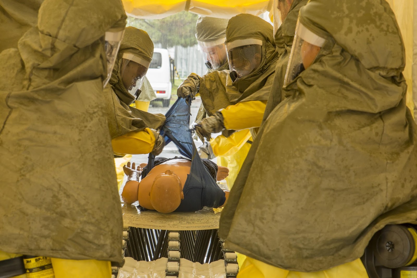
La gestión adecuada de los cadáveres es uno de los aspectos más complejos en la respuesta a las situaciones de desastre. Los desastres causan miles de muertes a nivel mundial cada año; sin embargo, no se le da atención al cuidado de los fallecidos en las actividades de planificación y la falta de guías para los primeros en responder se ha puesto de relieve después de varias grandes catástrofes. Esta guía de campo para personal no especializado ofrece orientaciones prácticas que facilitarán la recuperación, identificación básica, almacenamiento, la disposición, y en conjunto, la gestión adecuada de los cadáveres después de los desastres. También hace sugerencias sobre la forma de brindar ayuda a los familiares y de comunicarse con el público en general y con los medios de comunicación.
Este manual será de ayuda durante la respuesta inmediata a un desastre cuando aún no se cuenta con ayuda forense. Además podrá ser usado en la preparación de planes de desastres para el manejo de víctimas en masa. Las recomendaciones son relevantes para autoridades locales, regionales y nacionales, además de organizaciones no gubernamentales. Los principios y directrices enunciados en este documento ya se están ejecutando y promoviendo por varias organizaciones internacionales, incluidas las que han patrocinado la publicación del mismo: la Organización Panamericana de la Salud, la Organización Mundial de la Salud, el Comité Internacional de la Cruz Roja y la Federación Internacional de las Sociedades de la Cruz Roja y la Media Luna Roja.
El manual fue extensamente revisado por un grupo de expertos en el tema. Recibimos comentarios de ocho revisores técnicos: el dirigente del comité DVI de INTERPOL, el patólogo forense principal del Home Office en el Reino Unido, un especialista en medicina forense de Sri Lanka, dos administradores de desastres del Caribe, un acádemico experto en desastres del Reino Unido, un especialista en derechos humanos del Comité Internacional de la Cruz Roja (CICR) y un profesional internacional de desastres. Además, el manual fue revisado por los participantes de una reunión de especialistas forenses en Colombia, 15 líderes en salud pública de nueve países asiáticos en una reunión regional sobre el manejo de víctimas en masa, y expertos de medicina forense de Jordania que participaron en un taller del CICR. Asimismo, la versión preliminar del manual fue usada en el campo después del terremoto en Pakistán en 2005 y los deslizamientos de lodo en las Filipinas el mismo año.
La nueva edición en inglés refleja avances científicos y técnicos en el campo de gestión de víctimas en masa, y lecciones aprendidas del uso del manual.
-----------------------------------------------------------
Sigue este Blog en Facebook y Twitter
Este manual será de ayuda durante la respuesta inmediata a un desastre cuando aún no se cuenta con ayuda forense. Además podrá ser usado en la preparación de planes de desastres para el manejo de víctimas en masa. Las recomendaciones son relevantes para autoridades locales, regionales y nacionales, además de organizaciones no gubernamentales. Los principios y directrices enunciados en este documento ya se están ejecutando y promoviendo por varias organizaciones internacionales, incluidas las que han patrocinado la publicación del mismo: la Organización Panamericana de la Salud, la Organización Mundial de la Salud, el Comité Internacional de la Cruz Roja y la Federación Internacional de las Sociedades de la Cruz Roja y la Media Luna Roja.
El manual fue extensamente revisado por un grupo de expertos en el tema. Recibimos comentarios de ocho revisores técnicos: el dirigente del comité DVI de INTERPOL, el patólogo forense principal del Home Office en el Reino Unido, un especialista en medicina forense de Sri Lanka, dos administradores de desastres del Caribe, un acádemico experto en desastres del Reino Unido, un especialista en derechos humanos del Comité Internacional de la Cruz Roja (CICR) y un profesional internacional de desastres. Además, el manual fue revisado por los participantes de una reunión de especialistas forenses en Colombia, 15 líderes en salud pública de nueve países asiáticos en una reunión regional sobre el manejo de víctimas en masa, y expertos de medicina forense de Jordania que participaron en un taller del CICR. Asimismo, la versión preliminar del manual fue usada en el campo después del terremoto en Pakistán en 2005 y los deslizamientos de lodo en las Filipinas el mismo año.
La nueva edición en inglés refleja avances científicos y técnicos en el campo de gestión de víctimas en masa, y lecciones aprendidas del uso del manual.
- Descargue la nueva versión en inglés
- Descargue el documento en español (761.17 kB)
- Gestion des dépouilles mortelles lors de catastrophes - Télécharger la version française
- Descargue la versión en japonés
-----------------------------------------------------------
Sigue este Blog en Facebook y Twitter
lunes, 9 de octubre de 2017
Virus detection in Human and Other Primate Cell Lines
The high prevalence of contaminated cell cultures suggests that viral contaminations might be distributed among cultures. We investigated more than 460 primate cell lines for Epstein-Barr (EBV), hepatitis B (HBV), hepatitis C (HCV), human immunodeficiency virus type 1 (HIV-1), human T-cell leukemia/lymphoma virus I and II (HTLV-I/-II), and squirrel monkey retrovirus (SMRV) infections for risk assessment. None of the cell lines were infected with HCV, HIV-1, or HTLV-I/-II. However, one cell line displayed reverse transcriptase activity. Thirty-nine cell lines harbored EBV DNA sequences. Studies on the lytic phase of EBV revealed that five cell lines produce EBV particles and six further cell lines produced EBV upon stimulation. One cell line contained an integrated HBV genome fragment but showed no virus production. Six cell lines were SMRV-infected. Newly established cell lines should be tested for EBV infections to detect B-lymphoblastoid cell lines (B-LCL). B-LCLs established with EBV from cell line B95-8 should be tested for SMRV infections.
-----------------------------------------------------------
Sigue este Blog en Facebook y Twitter
REFERENCE:
Uphoff, Cord C. et al. “Detection of EBV, HBV, HCV, HIV-1, HTLV-I and -II, and SMRV in Human and Other Primate Cell Lines.” Journal of Biomedicine and Biotechnology 2010 (2010): 904767. PMC. Web. 8 Sept. 2017.-----------------------------------------------------------
Sigue este Blog en Facebook y Twitter
jueves, 5 de octubre de 2017
The Effects of Workplace Hazards on Female Reproductive Health
Many factors can affect a woman’s reproductive health and her ability to produce healthy children. We know that the health of an unborn child can suffer if a woman fails to eat right, smokes, or drinks alcohol during pregnancy. However, we know very little about the cause of most reproductive health problems such as infertility, miscarriage, and birth defects. We do know that some workplace hazards can affect a woman’s reproductive health, her ability to become pregnant, or the health of her unborn children. This document answers the following questions:
The Effects of Workplace Hazards on Female Reproductive Health. National Institute for Occupational Safety and Health. DHHS (NIOSH) Publication No. 99–104.
-----------------------------------------------------------
Sigue este Blog en Facebook y Twitter
- What are reproductive hazards for female workers?
- How does the female reproductive system work?
- What reproductive problems might be caused by workplace exposures?
- How are workers and their babies exposed?
- How are families exposed?
- How can exposures be prevented?
- What additional information is available?
The Effects of Workplace Hazards on Female Reproductive Health. National Institute for Occupational Safety and Health. DHHS (NIOSH) Publication No. 99–104.
-----------------------------------------------------------
Sigue este Blog en Facebook y Twitter
miércoles, 4 de octubre de 2017
Hospital Decontamination Self-Assessment Tool
In 2011, through a contract with the Massachusetts Department of Public Health, the Harvard School of Public Health Emergency Preparedness and Response Exercise Program (HSPH EPREP) engaged Massachusetts’ hospitals in a series of regional tabletop exercises focused on response to a hazardous materials incident. The exercise series highlighted a significant degree of heterogeneity among hospital decontamination programs and capabilities. Subsequent on-site assessments of hospital decontamination systems conducted at a representative sample of facilities throughout the Commonwealth confirmed this finding. To begin to address this issue of heterogeneity, HSPH-EPREP developed structured tools and guides to assist hospitals develop, maintain, and augment their decontamination programs. The Hospital Decontamination Self Assessment Tool was developed to provide hospitals with a means of evaluating decontamination plans and capabilities against current regulatory standards, recommendations from subject matter experts, and national and international healthcare decontamination best practices. This tool provides scalable considerations based upon presently available guidance to assist hospitals plan for and respond to small and large-scale incidents requiring the decontamination of patients contaminated by and/or exposed to chemical, biological, radiological, and/or nuclear agents.
REFERENCE:
Hospital Decontamination Self-Assessment Tool. A resource to assist hospitals evaluate decontamination plans and capabilities. Harvard School of Public Health. 2014
-----------------------------------------------------------
Sigue este Blog en Facebook y Twitter
REFERENCE:
Hospital Decontamination Self-Assessment Tool. A resource to assist hospitals evaluate decontamination plans and capabilities. Harvard School of Public Health. 2014
-----------------------------------------------------------
Sigue este Blog en Facebook y Twitter
martes, 3 de octubre de 2017
Best Practices for Hospital-Based First Receivers
Employers are responsible for providing a safe and healthful workplace for their employees. OSHA’s role is to assure the safety and health of America’s workers by setting and enforcing standards; providing training, outreach and education; establishing partnerships; and encouraging continual improvement in workplace safety and health. This handbook provides a general overview of a particular topic related to OSHA standards. It does not alter or determine compliance responsibilities in OSHA standards or the Occupational Safety and Health Act of 1970. Because interpretations and enforcement policy may change over time, you should consult current OSHA administrative interpretations and decisions by the Occupational Safety and Health Review Commission and the Courts for additional guidance on OSHA compliance requirements.
REFERENCE:
Best Practices for Hospital-Based First Receivers of Victims from Mass Casualty Incidents Involving the Release of Hazardous Substances. Occupational Safety and Health Administration. U.S. Department of Labor. OSHA 3249-08N. 2005
-----------------------------------------------------------
Sigue este Blog en Facebook y Twitter
lunes, 2 de octubre de 2017
INFECTION RISKS to new and expectant mothers in the workplace
Pregnancy is part of everyday life.
Being pregnant does not mean that you are
ill, but some infections, if they are
contracted in pregnancy, can affect the
health of the mother and baby. In very rare
cases, the baby may suffer serious harm and
this may result in permanent disability or
even death. This publication is a guide for employers on protecting the health and safety of employees who are new or expectant mothers. It deals with the risk of infections in the workplace.
REFERENCE:
Advisory Committee on Dangerous Pathogens. INFECTION RISKS to new and expectant mothers in the workplace. A guide for employers. HSE BOOKS.
-----------------------------------------------------------
Sigue este Blog en Facebook y Twitter
REFERENCE:
Advisory Committee on Dangerous Pathogens. INFECTION RISKS to new and expectant mothers in the workplace. A guide for employers. HSE BOOKS.
-----------------------------------------------------------
Sigue este Blog en Facebook y Twitter
jueves, 28 de septiembre de 2017
Herramientas para la gestión del riesgo químico
Este documento pretende dar a conocer algunos de los métodos y modelos que se encuentran disponibles, sus características básicas, las variables que consideran, la revisión bibliográfica de los mismos, el marco normativo en el que se desarrollan y ver dónde y cómo encajan estas herramientas en nuestro sistema de prevención de riesgos laborales, principalmente en el marco de la higiene industrial (ver figura 1) y en particular en la gestión de los riesgos derivados de la exposición a agentes químicos.
REFERENCIA:
HERRAMIENTAS PARA LA GESTIÓN DEL RIESGO QUÍMICO: Métodos de evaluación cualitativa y modelos de estimación de la exposición. Ministerio de empleo y seguridad social. Gobierno de España.
-----------------------------------------------------------
Sigue este Blog en Facebook y Twitter
REFERENCIA:
HERRAMIENTAS PARA LA GESTIÓN DEL RIESGO QUÍMICO: Métodos de evaluación cualitativa y modelos de estimación de la exposición. Ministerio de empleo y seguridad social. Gobierno de España.
-----------------------------------------------------------
Sigue este Blog en Facebook y Twitter
lunes, 25 de septiembre de 2017
Recomendaciones del @CDC en caso de terremotos. #fuertemexico #fuerzamexico
El sobrevivir a un terremoto y reducir el impacto en la salud requiere de preparación, planificación y práctica. Con mucha anticipación, usted puede acumular suministros de emergencia, identificar y disminuir los posibles riesgos en su hogar y practicar lo que debe hacer durante y después de un terremoto. Aprender qué medidas se deben tomar puede ayudarle a usted y a su familia a permanecer sanos y salvos en caso de un terremoto.
Esta guía contiene información general de que hacer antes, durante y después de un terremoto.
https://www.cdc.gov/es/disasters/earthquakes/index.html
-----------------------------------------------------------
Sigue este Blog en Facebook y Twitter
Esta guía contiene información general de que hacer antes, durante y después de un terremoto.
- Estar preparados
- Suministros de emergencia
- Riesgos en el hogar
- Seguridad en ambientes abiertos y cerrados
- Almacenamiento y purificación de agua de emergencia
https://www.cdc.gov/es/disasters/earthquakes/index.html
-----------------------------------------------------------
Sigue este Blog en Facebook y Twitter
Unintended spread of a #biosafety level 2 recombinant retrovirus
Background: Contamination of vertebrate cell lines with animal retroviruses has been documented repeatedly before. Although such viral contaminants can be easily identified with high sensitivity by PCR, it is impossible to screen for all potential contaminants. Therefore, we explored two novel methods to identify viral contaminations in cell lines without prior knowledge of the kind of contaminant.
Results: The first hint for the presence of contaminating retroviruses in one of our cell lines was obtained by electron microscopy of exosome-like vesicles released from the supernatants of transfected 293T cells. Random amplification of particle associated RNAs (PAN-PCR) from supernatant of contaminated 293T cells and sequencing of the amplicons revealed several nucleotide sequences showing highest similarity to either murine leukemia virus (MuLV) or squirrel monkey retrovirus (SMRV). Subsequent mass spectrometry analysis confirmed our findings, since we could identify several peptide sequences originating from monkey and murine retroviral proteins. Quantitative PCRs were established for both viruses to test currently cultured cell lines as well as liquid nitrogen frozen cell stocks. Gene fragments for both viruses could be detected in a broad range of permissive cell lines from multiple species. Furthermore, experimental infections of cells negative for these viruses showed that both viruses replicate rapidly to high loads. We decided to further analyze the genomic sequence of the MuLV-like contaminant virus. Surprisingly it was neither identical to MuLV nor to the novel xenotropic MuLV related retrovirus (XMRV) but showed 99% identity to a synthetic retrovirus which was engineered in the 1980s.
Conclusion: The high degree of nucleotide identity suggests unintended spread of a biosafety level 2 recombinant virus, which could also affect the risk assessment of gene-modified organisms released from contaminated cell cultures. The study further indicates that both mass spectrometry and PAN-PCR are powerful methods to identify viral contaminations in cell lines without prior knowledge of the kind of contaminant. Both methods might be useful tools for testing cell lines before using them for critical purposes.
REFERENCE:
Stang, Alexander et al. “Unintended Spread of a Biosafety Level 2 Recombinant Retrovirus.” Retrovirology 6 (2009): 86. PMC. Web. 8 Sept. 2017.
https://www.ncbi.nlm.nih.gov/pmc/articles/PMC2760500/
-----------------------------------------------------------
Sigue este Blog en Facebook y Twitter
jueves, 21 de septiembre de 2017
MAPA Información Centros de Acopio, derrumbes, hospitales y voluntariado #FuerzaMexico
Información general CDMX: http://cdmx.gob.mx/
Albergues CDMX:
http://www.cdmx.gob.mx/comunicacion/nota/listado-de-albergues-cdmx
Centros de Acopio CDMX
http://www.cdmx.gob.mx/comunicacion/nota/centros-de-acopio-gobierno-de-cdmx
MAPA
-----------------------------------------------------------
Sigue este Blog en Facebook y Twitter
Albergues CDMX:
http://www.cdmx.gob.mx/comunicacion/nota/listado-de-albergues-cdmx
Centros de Acopio CDMX
http://www.cdmx.gob.mx/comunicacion/nota/centros-de-acopio-gobierno-de-cdmx
MAPA
-----------------------------------------------------------
Sigue este Blog en Facebook y Twitter
lunes, 18 de septiembre de 2017
#WebinarAMEXBIO Transporte de Sustancias Infecciosas entre Instituciones
Registro
por internet en:
-----------------------------------------------------------
Detection of viral proteins in human cells lines by xeno-proteomics
Cell cultures used routinely in proteomic experiments may contain proteins from other species because of infection, transfection or just contamination. Since infection or contamination may affect the results of a biological experiment, it is important to test the samples for the presence of “alien” proteins. Usually cells are tested only for the most common infections, and most of the existing tests are targeting specific contaminations. Here we describe a three-step procedure for reliable untargeted detection of viral proteins using proteomics data, and recommend this or similar procedure to be applied to every proteomics dataset submitted for publication.
REFERENCE:
Chernobrovkin AL, Zubarev RA. Detection of viral proteins in human cells lines by xeno-proteomics: elimination of the last valid excuse for not testing every cellular proteome dataset for viral proteins. PLoS One. 2014 Mar 11;9(3):e91433. doi: 10.1371/journal.pone.0091433. eCollection 2014. PubMed PMID: 24618588; PubMed Central PMCID: PMC3950186
-----------------------------------------------------------
Sigue este Blog en Facebook y Twitter
miércoles, 13 de septiembre de 2017
Selecting Protective Clothing for Protection against Microorganisms in Blood and Body Fluids
Healthcare workers can be exposed to biological fluids that are capable of transmitting diseases. Those diseases, which are caused by a variety of microorganisms such as, Hepatitis B virus (HBV), Hepatitis C virus (HCV), Ebola Virus, and Human Immunodeficiency Virus (HIV) can pose significant risks to life and health. Healthcare workers wear protective clothing (e.g., surgical gowns, isolation gowns, and coveralls) to protect both patients and themselves from the transfer of microorganisms by blood and body fluids. A common misunderstanding among many end-users is that they are protected from blood, body fluids, and other potentially infectious materials when they wear any type of fluid-resistant garment. This document provides an overview of scientific evidence and information on national and international standards, test methods, and specifications for fluid-resistant and impermeable gowns and coveralls used in healthcare. This document focuses on selecting protective clothing primarily on the basis of their barrier properties; it does not address all aspects of garments related to their design, integrity, durability, comfort, and functionality.
REFERENCE
Considerations for Selecting Protective Clothing used in Healthcare for Protection against Microorganisms in Blood and Body Fluids
Download PDF HERE or HERE
-----------------------------------------------------------
Sigue este Blog en Facebook y Twitter
REFERENCE
Considerations for Selecting Protective Clothing used in Healthcare for Protection against Microorganisms in Blood and Body Fluids
Download PDF HERE or HERE
-----------------------------------------------------------
Sigue este Blog en Facebook y Twitter
lunes, 11 de septiembre de 2017
Guidance for Donning and Doffing Personal Protective Equipment (PPE) for #Ebola
The following informational materials demonstrate the procedures described in CDC guidance for donning and doffing (i.e., putting on and removing) personal protective equipment (PPE) for all healthcare providers entering the room of a patient hospitalized with known or suspected Ebola virus disease (Ebola). These informational materials are intended to promote patient safety and increase the safety of the healthcare provider.
Prior to working with Ebola patients, all healthcare providers involved in the care of Ebola patients must receive training and demonstrate competency in performing all Ebola-related infection control practices and procedures, specifically in donning and doffing proper PPE.
REFERENCE:
Guidance for Donning and Doffing Personal Protective Equipment (PPE) During Management of Patients with Ebola Virus Disease in U.S. Hospitals. CDC 2014.
-----------------------------------------------------------
Sigue este Blog en Facebook y Twitter
Prior to working with Ebola patients, all healthcare providers involved in the care of Ebola patients must receive training and demonstrate competency in performing all Ebola-related infection control practices and procedures, specifically in donning and doffing proper PPE.
REFERENCE:
Guidance for Donning and Doffing Personal Protective Equipment (PPE) During Management of Patients with Ebola Virus Disease in U.S. Hospitals. CDC 2014.
Sigue este Blog en Facebook y Twitter
jueves, 7 de septiembre de 2017
Acerca de las batas médicas
Las batas son ejemplos de equipos de protección personal utilizados en los entornos de atención médica. Se utilizan para proteger al usuario de la propagación de una infección o enfermedad si el usuario entra en contacto con material líquido y sólido potencialmente infeccioso. También pueden usarse para ayudar a evitar que el usuario del vestido pueda contaminar a pacientes vulnerables, como aquellos con sistemas inmunológicos debilitados. Las batas son una parte de una estrategia de control de infecciones. Algunos de los muchos términos que se han utilizado para referirse a las batas destinados a ser utilizados en los entornos de atención médica, incluyen batas quirúrgicas, ropa de aislamiento, ropa de aislamiento quirúrgico, batas no quirúrgicos, batas de procedimiento y batas sala de operaciones.
En 2004, la FDA reconoció el estándar de consensodel American National Standards Institute/Association of the Advancement of Medical Instrumentation (ANSI/AAMI) PB70:2003, “Liquid barrier performance and classification of protective apparel and drapes intended for use in health care facilities.”. La nueva terminología de la norma describe los niveles de protección de barrera de batas y otras prendas de protección destinadas a ser utilizadas en instalaciones de atención médica y especifica métodos de prueba y resultados de desempeño necesarios para verificar y validar los niveles de protección recientemente definidos:
- Nivel 1: Riesgo mínimo, para ser utilizado, por ejemplo, durante la atención básica, aislamiento estándar, vestido de cubierta para los visitantes, o en una unidad médica estándar
- Nivel 2: Bajo riesgo, que se utilizará, por ejemplo, durante la extracción de sangre, sutura, en la Unidad de Cuidados Intensivos (UCI), o un laboratorio de patología
- Nivel 3: Riesgo moderado, que se utilizará, por ejemplo, durante la extracción de sangre arterial, inserción de una vía intravenosa (IV), en la sala de emergencias o en casos de traumatismos
- Nivel 4: Alto riesgo, para ser utilizado, por ejemplo, durante largos procedimientos intensivos en líquidos, cirugía, cuando se necesita resistencia a patógenos o se sospecha de enfermedades infecciosas (no aerotransportadas)
- Medical gown. FDA
- ANSI/AAMI Standard PB70:2012 Liquid barrier performance and classification of protective apparel and drapes intended for use in health care facilities
- Premarket Notification Requirements Concerning Gowns Intended for Use in Health Care Settings
- Considerations for Selecting Protective Clothing used in Healthcare for Protection against Microorganisms in Blood and Body Fluids
Sigue este Blog en Facebook y Twitter
Suscribirse a:
Entradas (Atom)


